Viktor Simmons · 22 October 2026
Chemical Profiles of Kava Extracts and Their Measured Interactions with Human Liver Enzymes in Lab Settings
Kava extracts derived from Piper methysticum roots contain a range of kavalactones including kavain, dihydrokavain, methysticin, dihydromethysticin, yangonin, and desmethoxyyangonin, while additional compounds such as flavokavains A, B, and C appear in varying concentrations depending on extraction methods and plant cultivars, and laboratory analyses conducted through HPLC and GC-MS protocols have quantified these profiles across multiple commercial preparations since the early 2000s.
Those who study kava chemistry note that root powders and solvent extracts differ significantly in total kavalactone content, with aqueous preparations typically yielding lower percentages compared to ethanol or acetone based products, whereas regional varieties from Vanuatu and Fiji demonstrate distinct ratios that researchers track through standardized reference materials.
Standardized Extraction Methods and Resulting Profiles
Lab teams prepare kava extracts using solvents that range from water and ethanol to supercritical CO2, and each approach alters the final chemical makeup because polar solvents pull more hydrophilic components while nonpolar ones concentrate lipophilic kavalactones, so data from controlled experiments show total kavalactone levels spanning 5 to 70 percent by weight across samples tested in 2024 through 2026.
One study tracked seasonal harvests and found that noble cultivars maintain higher kavain percentages than tudei types, which contain elevated levels of dihydromethysticin, and these differences become measurable when extracts undergo fractionation followed by mass spectrometry confirmation.
Interactions Observed in Human Liver Microsome Assays
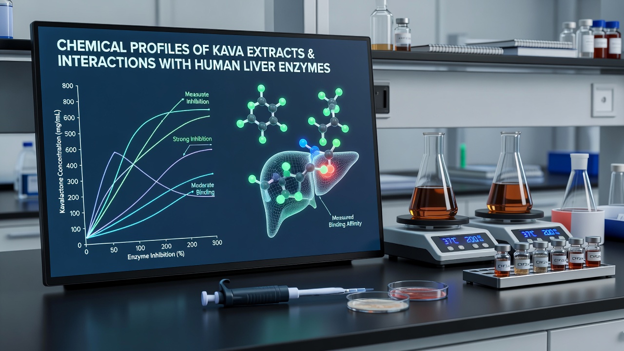
Researchers incubate kava extracts with human liver microsomes and recombinant CYP enzymes to measure inhibition constants, and results indicate that several kavalactones inhibit CYP3A4 and CYP2E1 at micromolar concentrations while flavokavain B shows activity against CYP2D6 and CYP1A2 in dose-dependent patterns documented across multiple publications.
Turnout from these assays reveals time-dependent inhibition for certain compounds, meaning preincubation with NADPH increases the extent of enzyme suppression, whereas reversible competitive inhibition occurs with others, and such distinctions matter because they help predict potential changes in drug metabolism rates when extracts reach therapeutic exposure levels.
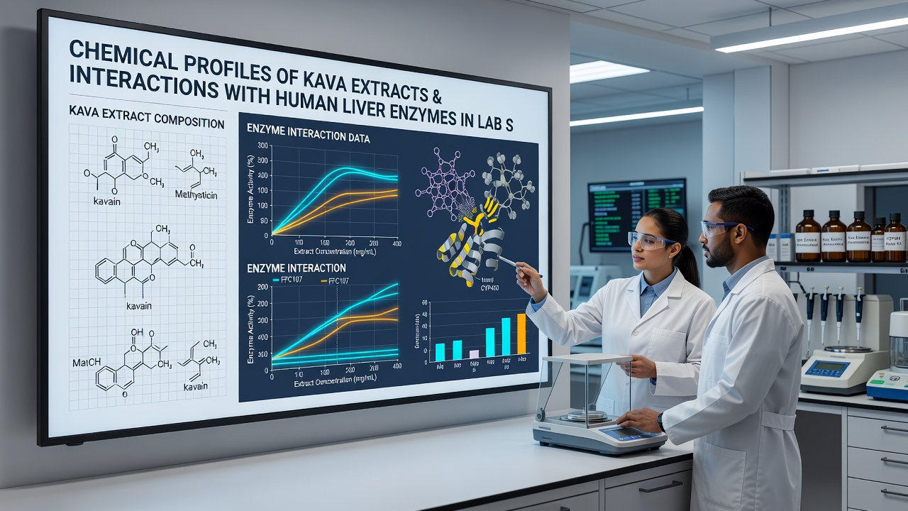
Additional experiments using cryopreserved human hepatocytes extend the microsomal findings by tracking parent compound depletion and metabolite formation over 24-hour periods, and data collected as recently as October 2026 confirm that yangonin undergoes rapid oxidative metabolism primarily through CYP3A4 pathways in these cellular models.
Key Enzyme Targets and Inhibition Constants
Quantitative measurements list IC50 values for major kavalactones against CYP3A4 between 5 and 40 micromolar, with methysticin and dihydromethysticin ranking among the stronger inhibitors in side-by-side comparisons, while CYP2C9 and CYP2C19 show milder responses that vary with extract concentration and co-incubated substrates such as midazolam or dextromethorphan.
Those running parallel screens note that whole extracts sometimes produce stronger inhibition than isolated compounds at equivalent doses, suggesting additive or synergistic effects among the kavalactone mixture, and this pattern holds across both recombinant enzyme systems and pooled human liver microsomes sourced from multiple donors.
Reports from the National Institutes of Health chemical genomics program document similar rank orders of potency, and corresponding work from Australian research groups using TGA-supported reference standards aligns closely with North American results on the same enzyme panel.
Metabolite Identification and Stability Data
LC-MS/MS methods identify hydroxylated and demethylated metabolites of kavain and yangonin formed during microsomal incubations, and these products accumulate at rates that depend on both extract type and added cofactors, while stability studies show that certain kavalactones degrade under prolonged light exposure or alkaline conditions before enzyme contact even begins.
Lab protocols now incorporate antioxidant stabilizers during extraction to preserve native profiles, and resulting data indicate that preserved extracts maintain consistent inhibition profiles across repeated freeze-thaw cycles in storage.
Conclusion
Lab-based measurements of kava extract chemistry and human liver enzyme interactions supply a growing dataset that regulatory and research communities continue to reference when evaluating extract standardization and potential metabolic interactions, and ongoing work through 2026 refines these profiles with higher-resolution instrumentation and broader donor panels for microsomal testing.